Sports and Energy Drink Consumption Damages Tooth Enamel
March 12th, 2026
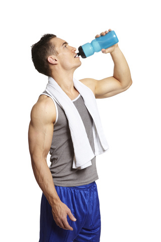
While they may sound refreshing, especially after participating in sports activities or after a jog, recent studies suggest that energy and sports drinks can damage tooth enamel, thus elevating your cavity risk. These drinks are especially popular among our younger patients.
In the study, researchers analyzed the fluoride content and pH levels of 13 sports and nine energy drinks by soaking tooth enamel samples in the aforementioned drinks. The samples were soaked for 15 minutes in each drink, and then were soaked for two hours in artificial saliva four times a day for five days.
As much as sports drinks are harmful to your teeth, researchers found that exposure to energy drinks such as Rockstar, Monster®, and Red Bull® resulted in twice as much enamel loss as exposure to sports drinks such as Powerade®, Gatorade®, and Propel® (3.1 percent to 1.5 percent).
Yes, there are health benefits to consuming orange juice, fruit juices, sports drinks, and flavored waters, which can contain valuable ingredients such as vitamin C and other antioxidants; these drinks can also replenish nutrients lost during a sporting event and lower the chance of heart disease and cancer. But, if not consumed carefully, these beverages can harm your teeth. They are full of sugar, which converts to acid and wears away at your teeth, causing cavities, sensitivity, and eventually tooth loss.
Even one drink a day is potentially harmful, but if you are absolutely unable to give up that sports or energy drink habit, we encourage you to minimize their use and rinse with water afterward or chew a sugar-free piece of gum. Do not brush immediately after drinking them; softened enamel due to acid is easier to damage, even when brushing. Remember, it takes your mouth approximately 30 minutes to bring its pH level back to normal. The best thing to do is to wait an hour, then brush to remove sugar that lingers on your teeth and gums.
There are many sports drinks, energy drinks, and flavored waters out there today. Dr. Keith J. Fabre Jr. and our team recommend you take the time to read the labels. Check for sugar content and citric acid in the ingredients. If you have any questions, or would like suggestions on the best sports drink options, please give us a call at our Marrero office or ask us during your next visit!